Top physicians,
technology and results stands behind our care for your health

Contact Us For a Free Assesment
Why Turan & Turan Health?

- Modern Technology
- Professional Doctors
- Focused solely on orthopedic conditions
- Our dedicated experts are available 24/7
- Personal assistance throughout your journey
- Completely free consultations
- Inexpensive while top-notch
Treatments
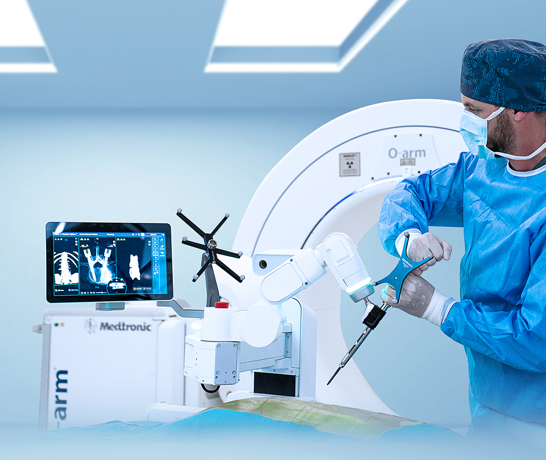
Our goal is to get you back to better. We offer a range of orthopedic services for common conditions and other diagnoses. Our expert physicians and therapists create customized treatment plans based on your goals – providing the advanced treatment and therapy you need. Come and see what makes Turan Turan Hospital different.
We Make It Easy
Medical Consultations
We offer online or in-person consultations with Turan&Turan orthopedic experts to help our patients determine the best treatment options for their needs.
Treatment Planning
We can help our patients plan their medical treatment in Turkey, including scheduling appointments, coordinating travel arrangements, and arranging transportation to and from medical facilities.
Accommodation Arrangements
We also can help patients find suitable accommodation options near the medical facility, such as hotels, apartments, or guesthouses.
Interpreter Services
Turan&Turan offers interpreter services to help our patients communicate with medical staff and navigate the local healthcare system.
Post-treatment Support
We offer post-treatment support services, such as follow-up appointments and physical therapy, to help our patients fully recover from their medical treatment.
Travel Assistance
Turan&Turan's expert patient representatives can provide assistance with travel-related issues, such as visa applications, travel insurance, and emergency medical assistance.
Stories
Reviews


News

Depression, Anxiety, and Substance Abuse on the Rise During the Pandemic
Originally coming from America, Black Friday is the day after Thanks giving which sees an impulsive sale across stores in preparation for Christmas. Consumers purchase


